To Improve the Quality of Tomosynthesis Images
| Subject: | 🏥 Health Care |
| Type: | Problem Solution Essay |
| Pages: | 5 |
| Word count: | 1446 |
| Topics: | Breast Cancer, Cancer, Computer Science, Disease, Innovation, Medicine, Nursing |

essay ASAP?
Table of Contents
Background
The increasing diagnosis of breast cancer in the US necessitates for a better clinically recognized modality for cancer detection to help in the reduction of mortality rate. Currently, mammography has been identified as the clinically accepted modality in the detection of breast cancer. However, mammography has numerous drawbacks that limits its specificity and sensitivity. A potential solution, therefore, endorses digital breast tomosynthesis (DBT) for inclusion.
Methods/Results
The methods used in this study aimed at;
- To develop a paired-image algorithm for the removal of artifact in the reconstruction of DBT.
- To improve the power spectrum estimator in the detectability calculation of the quality index of task based image through the modification of mammogram tube angulation.
- To increase the efficiency of dose through a reduction of random noise.
- To reduce the inefficiencies that are caused by less competent radiographer and wrong posture of the patient.
A paired-view method was used to develop a more efficient power spectrum estimator.


Conclusion/Implication
Apart from a reduced variance and bias in the estimation, the paired-view scheme in the reconstructed DBT had a better image quality as compared to the conventional average method. Hence, there was an improved finer frequency steps and better detectability in the measurement.
Problem
The current screen film mammography, which is highly recommended in the diagnosis and detection of breast cancer, is inherently partial for a number of reasons. First, the image is recorded in a film that has a fixed mapping of flounce by x-ray radiations due to its optical density, hence, the need for the incorporation of an external radiation to meet a certain level of image brightness (Chen, 2007). Second, the three dimensional arrangement of the breast is blurred by the two dimension projection of the image. Third, the granular structure in the film adds structural noise to the image. Fourth, there is a loss of the spatial resolution in the event where there is the thickening of the screen to absorb the x-ray due to the diffusion of light in the phosphor surface. Lastly, due to the nonlinear curve of the film surface, the image contrast is likely to vary with the film’s exposure, thus, inherent image is independent of the transmitted x-ray beam, which compromises the quality of the image (Chen, 2007). Therefore, a new technology that incorporates the impending improvement of the image quality and consequently, breast detection, only if properly used, is the adoption of digital breast tomosynthesis. Nonetheless, this study asserts that DBT inclusion alone does not guarantee an improved quality of tomosynthesis image, intrinsically, certain criteria have to be met for the method to give the best image. Therefore, the current task is aimed at improving the standard care for breast cancer patients by increasing patient safety, minimizing the amount of radiation exposure and provision of superior images. This study, consequently, seeks to find the underlying factors that would augment the functioning of DBT.


Background
Breast cancer is one of the most prevalent cancer in women globally. The American Cancer Society found that in 2010, approximately 207,000 new cases of cancer were recorded with close to 40,000 women to have succumbed to the disease (Philpotts & Hooley, 2016). The patient survival rate of five years can only be high when the cancer is diagnosed early enough and is quite low in late diagnosis, especially after the start of metastasis. However, despite the benefits of early diagnosis in lowering the mortality rate, yearly screening mammography is a vital option for women above 40 years. This has proven effective in reducing the rate at which breast cancer develops. Essentially, the application of tomosynthesis relies greatly on the strengths and ascertained abilities of mammography (Salkowski & Moseley, 2017). DBT has specifically been reported to have a lower x-ray projections that is able to improve the quality of the images in a pseudo three-dimensional tomographic imaging of the breast, which indicates the relevance for its adoption (Nelson et al., 2013).
Measurement
The data for the study was collected for six months. PGMI images were consequently graded based on the current standards applied for conventional mammography in NCCS (Sonnenschein & Waldherr, 2017). The project team keyed in the data for the low edge tomosynthesis paddle on a weekly basis, where modality leads and the deputy modality leads images were graded.
Design
The project team consisted of the project leader and five project members. In a Plan–Do–Study–Act (PDSA) cycle format, the team members were to begin by reviewing the key, in which they had to examine the RIS, then they were to select the correct patient folder from the mammography machine. The radiographer was consequently supposed to choose the correct views to perform the examination. This project used a low edge tomosynthesis paddle, which was mounted on the machine, in a correct position to get an exposure. The image was finally sent to PACS software for analysis as outlined in the flowchart below (Jiang et al., 2007).
Figure 1: 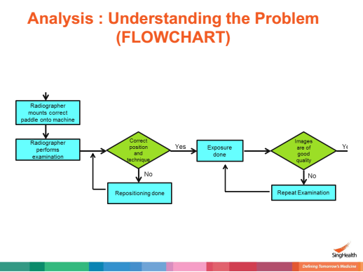
Strategy
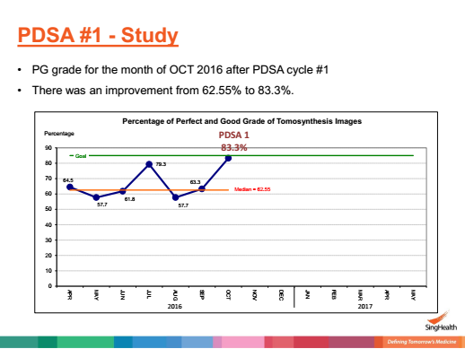
The initial PDSA cycle involved the planning, collection and keying of the tomosynthesis cases as well as the organization of the project with a single paddle. The team used only one low edge tomosynthesis paddle for numerous tomosynthesis cases, which was shared by two imaging rooms, lengthening the queuing time for patients. After the cycle, the team noted an improvement from 62.55% to 83.30% in the percentage of perfect and good grade of the image (figure 2). As such, the change of using low edge tomosynthesis paddle was implemented and the identified weakness was solved for the next cycle.
Figure 2: Percentage of Perfect and Good Grade of Tomosynthesis Images
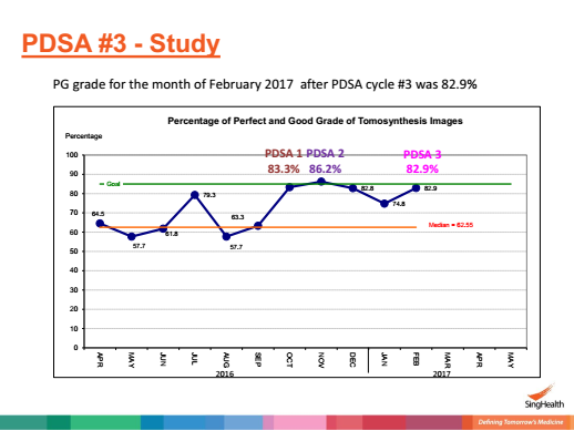
In the second PDSA cycle, all the factors were maintained from the first cycle, apart from the inclusion of a new low edge paddle, the modification of mammogram tube angulation and repositioning of the patient. This led to an improvement of the grade to 86.2% from 83.3% (figure 3), therefore, the changes were likewise implemented.
The third cycle was done after the training of the team members in positioning techniques and the grading of images. Therefore, since the result (82.9%) recorded a positive improvement from the median (62.5%), the team implemented the changes.
Results
The baseline data was used at the onset of the project to represent the contemporary mammography, which laid the foundation for the implementation of the changes. Subsequently, to determine the factors that would improve the quality of tomosynthesis images, certain alterations were made in PDSA. The first PDSA cycle recorded the greatest improvement in the quality of the image (62.55% – 83.30%) (figure 3); the inclusion of a low edge tomosynthesis paddle. Further improvement of the image with the addition of a new paddle led to an increase in the grade of the image from 83.3% to 86.2%, signifying the need for its implementation. Similarly, training of the radiographer (82.9%), improving the angulation of the mammogram tube and the repositioning of the patient had positive effects on the image grade, much far from the median grade.
Figure 3: Percentage of Perfect and Good Grade of Tomosynthesis Images
Lessons and Limitations
The study was projected to take place in six months (April to September), however, certain unanticipated occurrences delayed the completion of the final phase. At the start of the project, there was only one equipment to be used for multiple imaging, which delayed the process. Likewise, patients were positioned poorly and they were unable to endure the compression, causing a difficulty towards the achievement of a good image. The radiographers were also not familiar with the process, for instance, they had no log book to collect the cases, gave insufficient feedback, inexperienced, rushed and made mistakes. Therefore, this was a lesson to the project team to always plan and prepare for the anticipated challenges before the onset of the project. The team also learnt on the significance of incorporating experienced staff in the process, that is, training of staff to prepare them for the procedure would make the process flawless.
Conclusion
To improve the quality of tomosynthesis images, a new constrained paired-view method was incorporated in the image reconstruction for DBT. This involved the reduction of reconstruction artefacts that spread out of the plane slices, hence here was the enhancement of the conspicuity of viable lesions, which led to improved detectability and less distortions. Similarly, to enhance the quality of the tomosynthesis images, the study considered the effectiveness of other factors to do with radiography, including the training of radiographers, right positioning of the patient and the modification of the angular direction of mammogram tube.

- Chen, Y. (2007). Digital breast tomosynthesis (DBT): A novel imaging technology to improve early breast cancer detection: implementation, comparison and optimization.
- Jiang, Y., Sahiner, B., Society of Photo-optical Instrumentation Engineers., & American Association of Physicists in Medicine. (2007). Medical imaging 2007: 21-22 February 2007, San Diego, California, USA. Bellingham, Wash: SPIE.
- Nelson, G. S., Fahrig, R., Napel, S., Pelc, N. J., & Stanford University. (2013). Optimization and evaluation of scanning-beam digital X-ray tomosynthesis for real-time 3D image guidance for lung tumor biopsies.
- Philpotts, L. E., & Hooley, R. J. (2016). Breast Tomosynthesis. Saintt Louis: Elsevier Health Sciences.
- Salkowski, L., & Moseley, T. W. (2017). Clinical breast tomosynthesis: A case-based approach to screening and diagnosis. New York: Thieme.
- Sonnenschein, M., & Waldherr, C. (2017). Atlas of breast tomosynthesis: Imaging findings and image-guided interventions. Cham, Switzerland: Springer.

Offered for reference purposes only.